Crohn's Disease Pictures
Article
2025-12-08 • 5 min read

Crohn's Disease Pictures

Crohn disease is a complex and often visual condition. Images that depict Crohn disease in its various forms can be powerful tools for patient education, medical training, and research communication. When used thoughtfully, Crohn disease pictures help peopl...

Crohn disease is a complex and often visual condition. Images that depict Crohn disease in its various forms can be powerful tools for patient education, medical training, and research communication. When used thoughtfully, Crohn disease pictures help people understand the disease’s patchy, transmural inflammation, its potential impact on different segments of the gut, and the way symptoms relate to what is seen during investigations. They can bridge the gap between clinical terminology and everyday understanding, supporting informed conversations among patients, families, and clinicians.

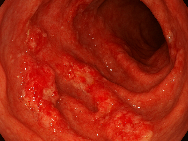
Images of Crohn disease appear across multiple modalities, each offering a different lens on the condition. Endoscopic pictures from colonoscopy or capsule endoscopy reveal mucosal patterns such as patchy inflammation, ulcers, cobblestoning, and sometimes strictures. Radiologic images from CT enterography or MR enterography show the deeper, transmural nature of disease, including wall thickening, creeping fat, and fistula formation. Pathology slides illustrate histologic features like focal granulomas and chronic inflammatory infiltrates. Medical education galleries, patient information portals, and stock image libraries curate these visuals to serve distinct purposes: teaching, patient counseling, or marketing communications for clinics and pharmaceutical companies. The common thread is clarity, accuracy, and sensitivity to the patient experience.

For professionals and educators, choosing the right set of images matters as much as the text that accompanies them. High quality visuals should be representative, clearly annotated when used for teaching, and paired with context about what the viewer is seeing and why it matters. For patients, images should illuminate the disease in a respectful and non alarmist way, helping them recognize signs that might warrant medical review while avoiding sensational or misleading portrayals. In both cases the goal is to support understanding without misrepresentation or overgeneralization. This is especially important because Crohn disease can present with a wide spectrum of appearances depending on the location and extent of disease, the therapy in use, and individual variation.

When it comes to sourcing Crohn disease pictures, several reputable avenues exist, each with its own strengths and licensing considerations. A practical approach is to explore a mix of free educational resources and paid libraries to balance accessibility, accuracy, and image quality.

A few prominent options include Radiopaedia, Wikimedia Commons, and NIH MedlinePlus. Radiopaedia specializes in radiology and includes case based imagery from CT and MR enterography that demonstrates the disease in the small intestine and colon. The strength here is clinical relevance; the images are typically annotated by radiology professionals and linked to concise case descriptions. Licensing and access can vary, with many images available for educational use and some requiring subscription or attribution, so it is important to check the terms before reuse. Wikimedia Commons hosts a broad collection of images ranging from endoscopic stills to histology slides. Because licensing is varied, users can often download and reuse images with appropriate attribution under Creative Commons or public domain licenses. This makes Wikimedia a valuable resource for educators and patients seeking free materials, provided the licenses are respected. NIH MedlinePlus is a trusted patient education portal with curated medical images that explain conditions in accessible language. Its imagery is designed for lay audiences, making it well suited for patient handouts and clinic waiting room materials. Licensing tends toward broad educational use, but users should still verify how the images can be reused in their own materials.

Crohn's Disease Pictures

For those seeking high fidelity or physician oriented visuals, paid sources such as VisualDx or stock image libraries like Getty Images and Shutterstock offer professional photographs and illustrations. VisualDx emphasizes clinical accuracy and visual differential diagnosis, with images that can be paired with patient education content and clinical notes. Stock libraries provide a wide range of Crohn related imagery, including professional photography and stylized illustrations. The tradeoff is cost and licensing complexity, as commercial use usually requires a license, and it is essential to respect copyright and ensure that images reflect current medical understanding.

If your goal is to assemble a balanced set of Crohn disease pictures for a patient education guide, here is a practical workflow: - Define the educational objective: patient overview, clinician training, or research briefing. - Choose a mix of modalities: a colonoscopy or capsule endoscopy image for mucosal appearance, a radiology image for transmural involvement, and a patient friendly diagram for quick comprehension. - Check licensing and attribution: confirm whether images require attribution, license type, and any restrictions on redistribution or modification. - Prioritize accuracy and currency: prefer images from reputable medical sources or recently published materials to reflect current understanding of Crohn disease. - Add accessible captions and alt text: provide plain language explanations of what the viewer sees and why it matters, optimize for screen readers, and avoid overly technical jargon when used for patient materials. - Consider accessibility and inclusivity: use color palettes accessible to colorblind viewers, avoid sensational framing, and protect patient privacy in any real world imagery.

Beyond sourcing, the design and layout of Crohn disease visuals contribute to comprehension. Use clear labels to identify key features such as mucosal ulcers, skip lesions, fistulas, or stenosis. Maintain a consistent visual style across images to help learners compare findings across modalities. Where possible, include a short caption that explains the clinical relevance of the image, what the viewer should look for, and how it ties to symptoms or treatment decisions. For patient facing materials, pair images with brief plain language explanations and practical steps readers can take if they notice similar symptoms.

When working on content that includes Crohn disease pictures, it is prudent to respect patient dignity and privacy. Where real patient imagery is used, ensure consent and remove identifiers. Where possible, opt for anonymized, de-identified, or illustration based visuals to convey clinical concepts without compromising individual rights. Finally, provide guidance about limitations of images. A single picture cannot capture the full scope of Crohn disease activity or its dynamic response to therapy. Encourage readers to interpret visuals within the broader clinical context, including symptoms, history, lab data, and imaging results interpreted by qualified clinicians.

In summary, Crohn disease pictures are a powerful adjunct to words. They help explain the disease to patients, illustrate learning points for students, and support clinical communication. By selecting credible sources, respecting licensing, and pairing visuals with thoughtful captions and accessible design, educators can create materials that educate without confusing, inspire without sensationalism, and ultimately support better understanding and management of Crohn disease.

← Back to all articles